Understanding Jaw Deficiency
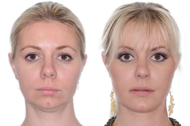
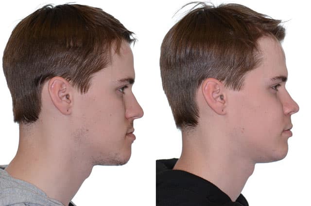
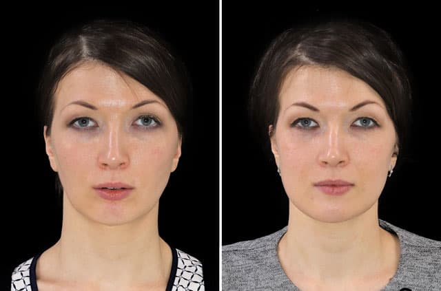
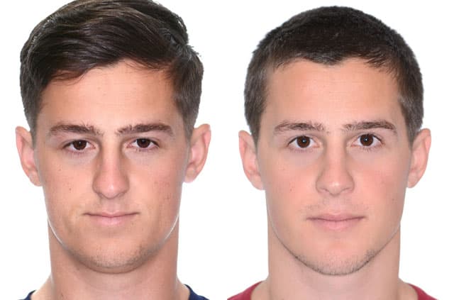
When both jaws are underdeveloped, the entire lower and middle face can appear set back, and the soft tissues lose the support they need to look full and balanced. This case involved a maxillary and mandibular deficiency classified as a Class III malocclusion, along with poor nasal and lip support. In simple terms, both the upper and lower jaws lacked adequate forward projection.
Deficient jaws can affect more than appearance. The space behind the tongue and soft palate may be narrowed, the lips and nose may sit without firm underlying support, and the bite often does not function ideally. Advancing both jaws forward is a well-established way to address these concerns by restoring projection to the facial skeleton and improving support for the surrounding soft tissues.
The Diagnosis Explained
The documented diagnosis included maxillary hypoplasia, meaning an underdeveloped upper jaw, and mandible deficiency, meaning the lower jaw was likewise lacking adequate development. Together these created a recessive overall facial structure that benefited from advancement of both jaws.
Poor nasal and lip support was also noted, reflecting how a deficient skeleton fails to prop up the overlying soft tissues. The case was further described as a maxillary and mandibular deficiency Class III malocclusion. Mapping these findings clarified that the goal was to bring both jaws forward to restore projection, support, and a more functional bite relationship.
The Surgical Plan
The upper jaw was addressed with a maxillary Le Fort I osteotomy performed as a 3-piece advancement of approximately 10mm, as documented. The 3-piece approach allows the surgeon to shape and widen the arch while advancing it, and a substantial advancement like this restores significant midface projection and improves support for the nose and lips.
Bone grafting to the maxilla was used to fill and stabilize the spaces created by advancing the jaw, promoting solid healing in the new position. The lower jaw was then brought forward with a mandibular bilateral sagittal split osteotomy advancement (BSSO), so that both jaws moved together — the essence of maxillomandibular advancement.
A genioplasty was performed to lengthen and set back the chin, fine-tuning the lower face after the jaws were advanced. Coordinating these procedures allowed the surgeon to balance projection across the midface, lower face, and chin while establishing a more functional bite.
Pre-Surgical Orthodontics and Treatment Planning
Maxillomandibular advancement for a Class III deficiency depends heavily on preparation. Most patients complete a phase of pre-surgical orthodontics, often lasting many months, during which braces or aligners level and coordinate each dental arch and remove the dental compensations that develop around deficient jaws. In a Class III pattern the lower teeth tend to tip back and the upper teeth flare forward to camouflage the imbalance, so reversing those compensations is essential for the advanced jaws to meet in a stable, functional bite.
Detailed three-dimensional planning determines how much projection to restore. Imaging, dental models, and facial analysis help the surgeon plan the roughly 10mm 3-piece Le Fort I advancement, identify where bone grafting will support the lengthened maxilla, set the amount of mandibular advancement with the bilateral sagittal split osteotomy, and refine the chin with genioplasty. Because the upper and lower jaws move forward together, this blueprint is what keeps the advancement balanced and improves support for the nose, lips, and the airway behind the tongue.
This sequence reflects the close partnership between the orthodontist and the oral and maxillofacial surgeon that defines orthognathic surgery and malocclusion correction. A final phase of orthodontics after surgery typically refines how the teeth settle together. For patients across Roseville, Sacramento, and Placer County, understanding this team-based timeline helps frame the full course of jaw advancement surgery from preparation through finishing.
Recovery and What to Expect
Recovery from maxillomandibular advancement with bone grafting generally follows a staged course. Swelling and bruising are typically greatest in the first one to two weeks and then improve steadily. A soft or liquid diet during early healing protects the advanced and grafted bone while it begins to stabilize.
Because bone grafting accompanied the advancement, the graft needs time to integrate and the jaws need time to consolidate, a process that continues over several months. Most patients find that visible swelling largely resolves within several weeks while deeper healing proceeds. Orthodontic treatment commonly works alongside the surgery to finalize the bite, and follow-up appointments help confirm stable healing. These describe general expectations rather than an individual guarantee.
Corrective Jaw Surgery in Roseville, CA
This maxillomandibular advancement was performed by Dr. Alexander V. Antipov at Galleria Oral & Facial Surgery in Roseville, CA. Advancing both jaws with a 3-piece Le Fort I osteotomy, bone grafting, BSSO, and genioplasty requires meaningful surgical experience and precise planning to deliver balanced, stable results.
Patients throughout Roseville, Sacramento, Placer County, and Northern California come to our practice for corrective jaw surgery to address recessive jaws, weak facial support, and bite problems. If you feel your jaws and chin look set back or your bite does not work the way it should, we invite you to schedule a consultation to learn whether jaw advancement surgery could help.