Understanding Maxillary Deficiency
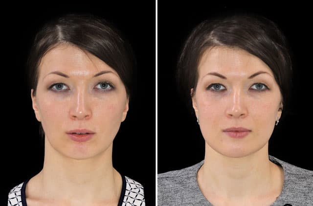
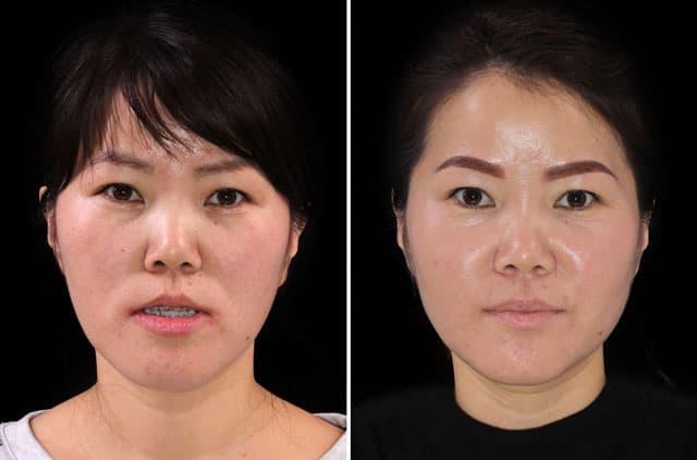
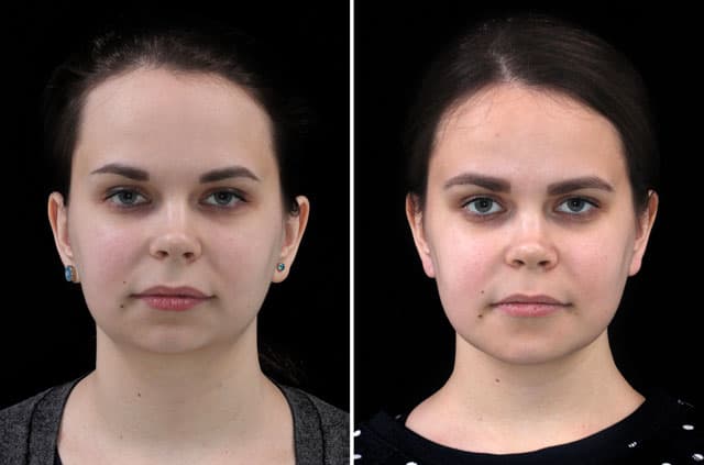
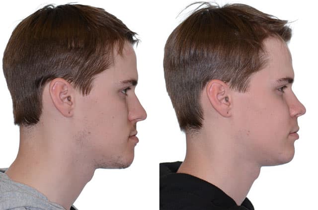
When the upper jaw is underdeveloped, the midface can appear flat or set back and the bite often falls into a Class III pattern. In this case, the patient had maxillary hypoplasia with a mandible of normal length, producing a Class III malocclusion. The additional findings of an inferior border chin deviation, a nasal deviation, and a left temporomandibular disorder rounded out the picture.
A deficient upper jaw with a normal lower jaw means the imbalance comes primarily from the maxilla sitting too far back. This relationship can affect how the teeth meet, how the midface looks, and how the surrounding features are supported. Targeted correction of the upper jaw is a logical, well-established way to restore projection and bring the bite into a healthier relationship.
The Diagnosis Explained
The documented findings centered on maxillary hypoplasia, an underdeveloped upper jaw, with the mandible noted as normal length. This combination is what produced the Class III malocclusion, a bite relationship in which the lower teeth sit forward of the upper teeth because the upper jaw is deficient rather than the lower jaw being excessive.
Additional findings included an inferior border chin deviation, meaning the bony lower edge of the chin was shifted off center, and a nasal deviation reflecting how skeletal imbalance can influence nasal appearance. A left temporomandibular disorder indicated symptoms in the left jaw joint. Documenting each of these elements helped focus the surgical plan on advancing the deficient upper jaw while accounting for the patient's overall presentation.
The Surgical Plan
The correction centered on a maxillary Le Fort I osteotomy performed as a one-piece procedure. In a Le Fort I osteotomy, the surgeon makes a precise horizontal cut that frees the upper jaw so it can be repositioned forward and leveled. Done as a single piece, the entire maxilla is moved as one unit to advance the deficient midface and improve the bite relationship.
Bone grafting to the maxilla was used to fill and stabilize the spaces created when the upper jaw was advanced. Grafting supports reliable healing by providing scaffolding for new bone to form in the gaps, helping the maxilla consolidate securely in its new, more forward position.
Focusing the correction on the upper jaw made sense given that the lower jaw was of normal length. By advancing the maxilla and supporting it with bone grafting, the plan aimed to restore midface projection, address the Class III relationship, and improve overall facial balance.
Pre-Surgical Orthodontics and Planning
Correcting a Class III malocclusion driven by maxillary hypoplasia begins with thorough preparation. Most patients complete a phase of pre-surgical orthodontics, often lasting many months, during which braces or aligners level and coordinate each dental arch and remove the dental compensations that develop around a deficient upper jaw. In a Class III pattern the teeth frequently tip to disguise the imbalance, so reversing those compensations is what allows the advanced maxilla to meet the lower teeth in a stable, functional bite.
Because the mandible was of normal length, planning focused on the upper jaw. Three-dimensional imaging, dental models, and facial analysis help the surgeon decide how far to advance and level the maxilla with the one-piece Le Fort I osteotomy and where bone grafting will be needed to stabilize the new position. Planning also accounts for the inferior border chin deviation, the nasal deviation, and the left temporomandibular disorder, so the maxillary advancement is positioned to improve facial balance while supporting comfortable jaw-joint function rather than straining it.
This sequence reflects the close partnership between the orthodontist and the oral and maxillofacial surgeon that defines orthognathic surgery and malocclusion correction. A final phase of orthodontics after surgery typically refines how the teeth settle together. For patients across Roseville, Sacramento, and Placer County, understanding this team-based timeline helps set realistic expectations for the full course of upper jaw advancement from preparation through finishing.
Recovery and What to Expect
Recovery after a Le Fort I osteotomy with bone grafting generally proceeds in stages. The first one to two weeks usually bring the most swelling and bruising, which then steadily subside. A soft or liquid diet during early healing protects the advanced and grafted upper jaw as it begins to stabilize.
Because bone grafting was part of this treatment, the graft needs time to integrate and the maxilla needs time to consolidate, a process that continues over several months. Most patients find that visible swelling largely resolves within several weeks while deeper healing proceeds quietly. Orthodontic treatment commonly complements the surgery to finalize the bite, and follow-up visits help confirm stable healing and a settled jaw position. These reflect general expectations for this procedure rather than a guarantee for any one patient.
Orthognathic Surgery in Roseville, CA
This correction was performed by Dr. Alexander V. Antipov at Galleria Oral & Facial Surgery in Roseville, CA. Advancing a deficient upper jaw with a Le Fort I osteotomy and bone grafting requires careful planning to restore both function and facial balance, especially when joint symptoms and asymmetry are also present.
Patients from Roseville, Sacramento, Placer County, and throughout Northern California turn to our practice for orthognathic surgery to correct jaw deficiencies, Class III bites, and facial imbalance. If you feel your midface looks flat, your lower teeth sit ahead of your upper teeth, or your jaw is uncomfortable, we encourage you to schedule a consultation so we can evaluate your situation and discuss your options.