Understanding Short Face Syndrome
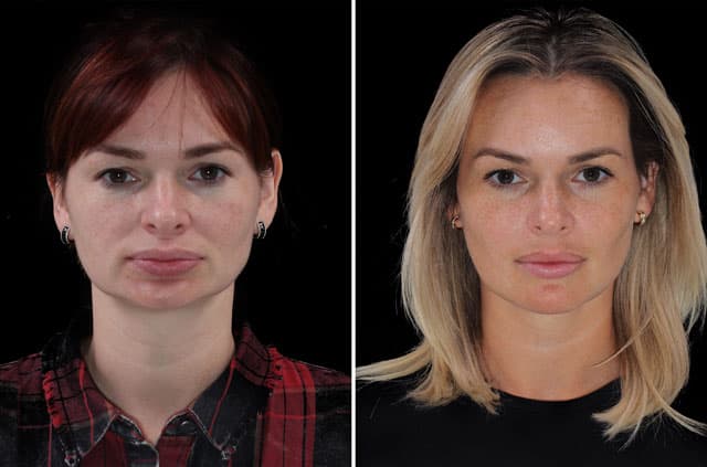
Short face syndrome describes a face in which the lower third appears compressed because the jaws have not developed enough vertical height. In this case, the patient had a Class I occlusion combined with reduced lower facial height — meaning the teeth met in a relatively normal class relationship, but the underlying jaws were too short, leaving the midface and lower face collapsed and the chin foreshortened.
As documented, the presentation included poor nasal and lip support, a short chin, and a collapsed midface and lower face. When the skeletal foundation lacks height, the soft tissues of the lips and nose lose their underlying support, and the smile can look cramped. Correcting this requires adding height and projection to the facial skeleton rather than simply treating the teeth, which is why a comprehensive surgical approach was planned.
The Diagnosis Explained
The documented findings paint a clear picture. Maxillary hypoplasia means the upper jaw was underdeveloped, and mandible deficiency means the lower jaw was also lacking adequate growth. Together these produced a short face and a short chin, with insufficient height across the lower portion of the face.
Additional findings included a collapsed midface and lower face and poor nasal and lip support, both consequences of a skeleton that did not provide enough underlying structure. The occlusion itself was classified as Class I malocclusion, indicating that while the bite category was relatively normal, the vertical and supportive deficiencies still warranted surgical correction. Identifying each of these elements ensured the plan would restore both height and projection where they were missing.
The Surgical Plan
The upper jaw was addressed with a 3-piece Le Fort I osteotomy and advancement. In a Le Fort I procedure, the maxilla is separated along a precise line so it can be repositioned; performing it in three pieces gives the surgeon additional control to widen and shape the arch as well as advance and lengthen it. This was key to restoring midface support and improving the smile.
Bone grafting to the maxilla was used to fill and stabilize the gaps created when the jaw was lengthened and advanced, supporting solid healing in the new, taller position. The lower jaw was then advanced with a bilateral sagittal split osteotomy (BSSO), bringing the mandible forward to balance the lengthened upper jaw and improve the profile and airway.
Finally, a genioplasty lengthened and advanced the chin to complete the vertical restoration of the lower face. Together, these procedures were designed to add the missing height and projection, improve nasal and lip support, and create more balanced facial proportions while maintaining a functional bite.
Pre-Surgical Orthodontics and Treatment Planning
Even when the occlusion is classified as Class I, correcting short face syndrome requires careful preparation so the vertical changes are stable. Most patients complete a phase of pre-surgical orthodontics in which braces or aligners level and coordinate each dental arch and remove any dental compensations that have developed around the underdeveloped jaws. Because the goal here is to lengthen the face rather than simply reclassify the bite, the orthodontic groundwork ensures the teeth still meet correctly once the maxilla and mandible are repositioned to a taller, more forward position.
Detailed three-dimensional planning guides how much height and projection to add. Imaging, dental models, and facial analysis help the surgeon decide how to expand and advance the upper jaw with the 3-piece Le Fort I osteotomy, where bone grafting will be needed to stabilize the lengthened maxilla, how far to advance the lower jaw with the bilateral sagittal split osteotomy, and how to lengthen the chin with genioplasty. This blueprint keeps the changes balanced so that nasal and lip support, midface fullness, and the smile all improve together.
This planning reflects the close collaboration between the orthodontist and the oral and maxillofacial surgeon that underpins orthognathic surgery and facial proportion correction. A final phase of orthodontics after surgery usually refines how the teeth settle. For patients across Roseville, Sacramento, and Placer County, understanding this team-based timeline helps set realistic expectations for the months of preparation and healing that surround a vertical lengthening procedure.
Recovery and What to Expect
Recovery after multi-level jaw surgery with bone grafting generally proceeds in phases. The first one to two weeks typically bring the most swelling and bruising, which then steadily improve. A soft or liquid diet is usually recommended early on to protect the grafted and repositioned bone as healing begins.
Because bone grafting was part of this treatment, the body needs time for the graft to integrate and the lengthened jaw to consolidate, a process that continues over several months. Most patients see the majority of visible swelling resolve within several weeks while deeper healing proceeds quietly underneath. Orthodontic treatment commonly complements the surgery to finalize the bite, and follow-up visits help confirm that healing is on track. These are general expectations for this procedure type rather than a guarantee for any one patient.
Corrective Jaw Surgery in Roseville, CA
This short face correction was performed by Dr. André-David Kahwach at Galleria Oral & Facial Surgery in Roseville, CA. Restoring vertical facial height with a 3-piece Le Fort I osteotomy, mandibular advancement, bone grafting, and genioplasty demands advanced surgical skill and detailed planning.
Our practice serves patients across Roseville, Sacramento, Placer County, and Northern California who are seeking improved facial proportions and bite function. If you feel your lower face looks short, your smile appears cramped, or your lips and nose lack support, a consultation can help determine whether corrective jaw surgery is a good fit. We welcome the chance to discuss your goals and explain the possibilities.